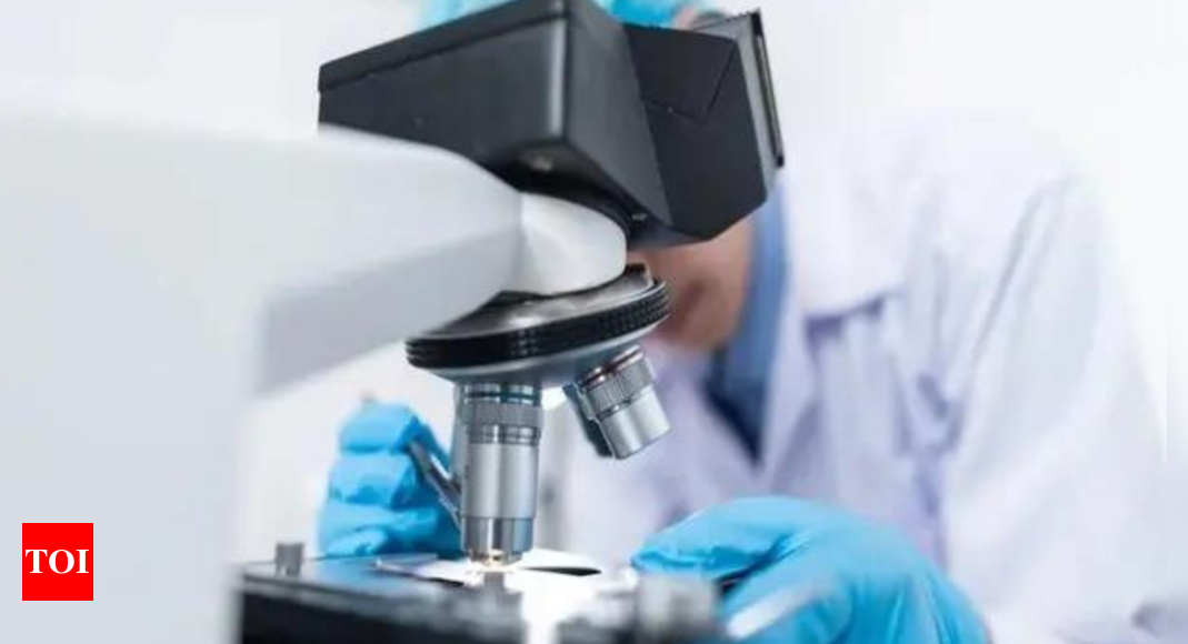
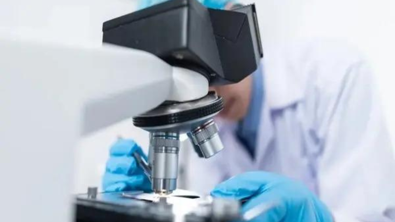
A new research has revealed that a common food dye, tartrazine (FD&C Yellow No. 5), can temporarily render the skin of living mice transparent, allowing for unprecedented visualization of internal structures without invasive procedures.
This groundbreaking study, published in the journal Science, marks a significant advancement in biomedical imaging techniques.This discovery, reminiscent of the serum in HG Wells’ novel “The Invisible Man,” allows scientists to observe the functioning of internal organs without invasive procedures, a CNN report said.
Mechanism of transparency
The transparency effect is achieved through a process that alters the refractive indices of the tissues involved. Biological tissues, such as skin and muscle, scatter light due to their heterogeneous composition, which includes proteins, fats, and liquids. This scattering is what typically prevents us from seeing through these tissues. The researchers discovered that by applying a concentrated solution of tartrazine to the skin, they could match the refractive indices of the various components within the tissue. This balancing act significantly reduces light scattering, allowing light to pass through more effectively, thus rendering the skin transparent.
When the tartrazine solution is applied, it absorbs certain wavelengths of light, particularly red light, which changes how light interacts with the tissue. For instance, the dye was shown to make the skin on the mice’s skulls and abdomens transparent, enabling researchers to observe blood vessels, internal organs, and even muscle contractions in real-time. The process is reversible; once the dye is rinsed off, the skin returns to its original opaque state.
Experimental findings
In the experiments, researchers first tested the dye on slices of chicken breast, which became transparent to red light after immersion in the tartrazine solution. Following these initial tests, they applied the dye to the scalps and abdomens of live mice. Within minutes, the researchers could visualize blood flow in the brain and identify organs such as the liver, small intestine, and bladder through the skin. The technique allowed for the observation of physiological processes, such as the beating heart and respiratory movements, providing a dynamic view of living tissues.
The application of the dye induced minimal inflammation and no long-term health effects were noted in the mice, as verified by subsequent blood tests and body weight measurements. The researchers concluded that this method could serve as a non-invasive tool for medical diagnostics, potentially allowing for the identification of tumors or injuries without the need for surgical intervention.
Potential applications
The implications of this research are vast. If proven safe for human use, this technique could revolutionize medical imaging and diagnostics. For example, it could simplify blood draws by making veins more visible, enhance the effectiveness of laser-based tattoo removal, and facilitate early cancer detection by allowing clinicians to examine tissues without invasive biopsies.
“For those who understand the fundamental physics behind this, it makes sense; but if you aren’t familiar with it, it looks like a magic trick,” said study author Zihao Ou. Co-author Guosong Hong added, “This innovation could assist in the early detection of skin cancer and make veins more visible.”
While the current study has not yet been tested on humans, the researchers are optimistic about the future applications of this technique. They acknowledge that human skin is thicker than that of mice, which may pose challenges for the absorption of the dye. Future studies may explore the use of microneedle patches or injections to facilitate deeper delivery of the dye, potentially overcoming this barrier.